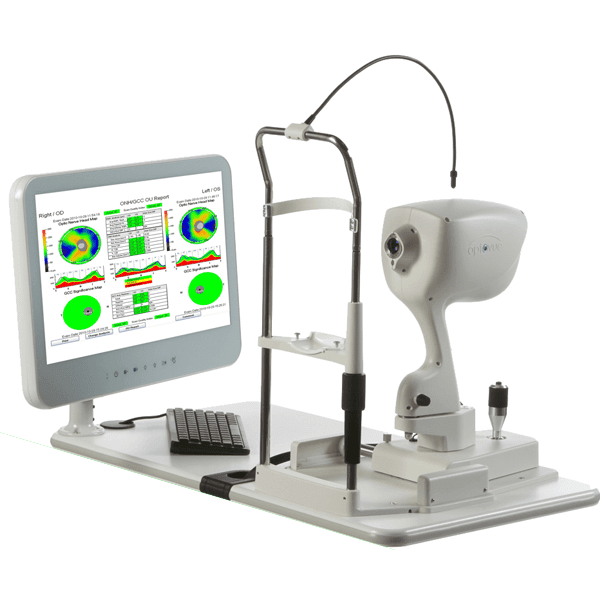
INTRODUCING THE OPTOVUE iVUE OCT
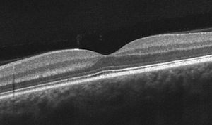
One of the most sophisticated instruments currently in eye care is the optical coherence tomographer (OCT). This technology uses light waves to create cross-sectional images of the retina and optic nerve. In other words, it allows us to analyze the microscopic tissues in the eye to detect various eye diseases!
These images are just like X-rays or MRIs. Sometimes we can see diseases without these scans, but these scans give us additional information that normally cannot be seen with traditional techniques. This technology is amazingly advanced and truly sets a new standard for eye care going forward.

WHAT DOES THE OCT DO?
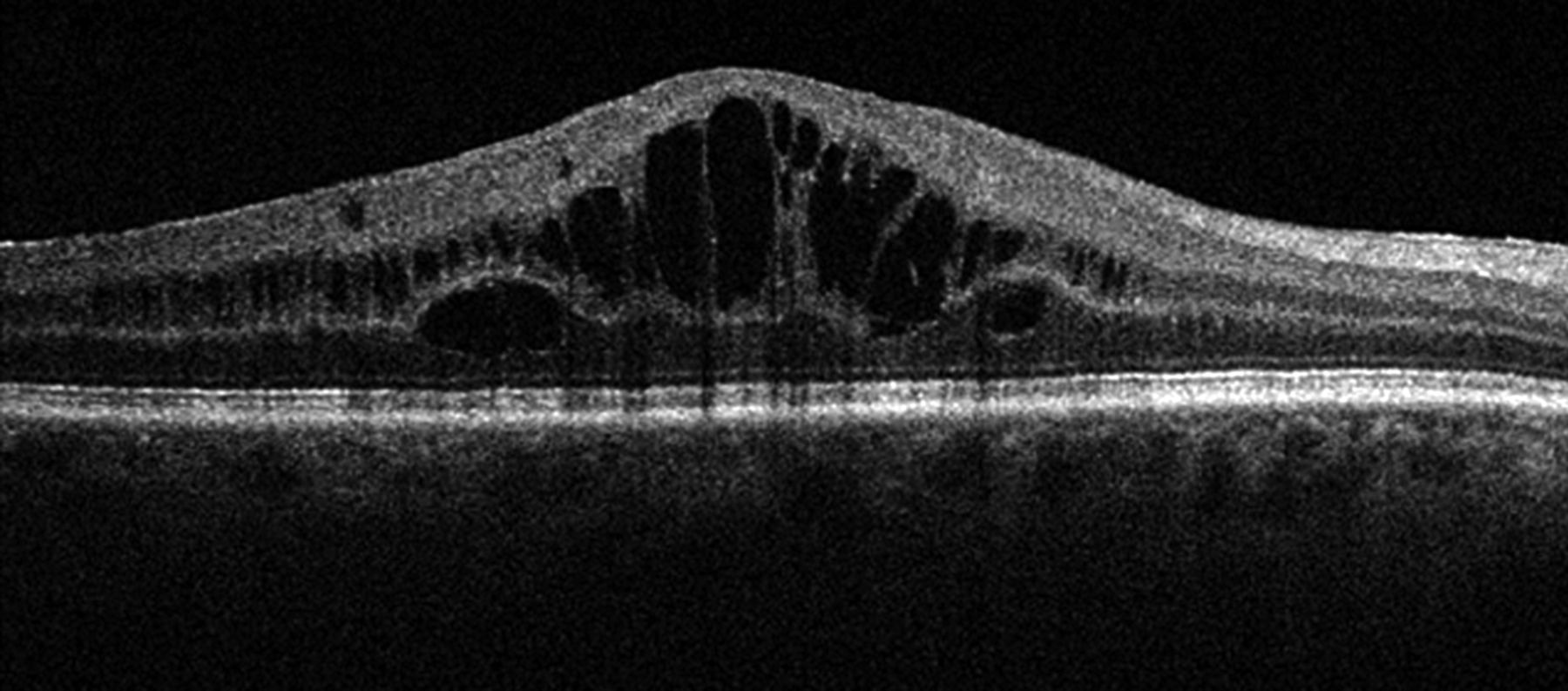
The OCT is a versatile piece tool with various functions. The primary use is for detecting diseases that affect the optic nerve and macula, two of the most important structures in the eye. For those of you that have had an exam with us in the past, our doctors have most likely showed you these structures in your fundus photos. The OCT allows us to see beyond the superficial surface of these structures to see what is happening underneath them. This is especially important for detecting and managing eye diseases such as macular degeneration, glaucoma, and diabetic retinopathy because these conditions often begin beneath the surface and cause microscopic changes that would otherwise be undetectable.
In addition to scanning the inner linings of the eye, the OCT can also scan the front surfaces to measure the thickness of your cornea and cells. It also helps us to ensure that contact lenses are fitted properly on the eye and allows us to make tiny adjustments that could greatly improve visual quality and comfort.


WHO SHOULD HAVE AN OCT SCAN?
OCT scans are a no-brainer if we are managing a patient for disease such as glaucoma, macular degeneration, or diabetes, but they are also excellent for detecting problems that may not be otherwise found with photos or dilated eye exams.
In glaucoma, the OCT provides us important information regarding the thickness of retinal and nerve tissues that allow us to see progressive thinning of those tissues, which may indicate the need for additional treatment. Retinal deposits and ruptured retinal tissues can be detected and monitored in worsening macular degeneration. For uncontrolled diabetics, we may see leaking blood vessels or swelling of the central retina.
In addition to these, many other conditions may be monitored and/or detected with the OCT including:
- Central serous chorioretinopathy
- Hypertensive retinopathy
- Optic nerve drusen
- Retinal artery and vein occlusions
- Toxicity caused by medications such as plaquenil
- Posterior vitreous detachments
- Macular pucker
- Macular holes
For patients without existing eye diseases, we encourage them to have an iWellness screening, which is an abbreviated scan that helps to quickly determine whether a patient's macula and optic nerve are suspicious for any conditions that need to be monitored. This scan is extremely useful for patients who may be at risk for certain eye conditions due to factors such as family history, medication use, or eye injuries.
Basically, all patients can benefit from this type of scan, and it is truly allowing our doctors to provide our patients with the best, most comprehensive eye care possible.
We are so excited to have this technology in our practice to provide our patients with the very best and most advanced eye care possible. No matter how complicated your case may be, Simply Optometry is here to simplify it for you.
